
Diagnosing and Treating Corneal Ulcers in Horses: Causes, Treatments, and Prognosis
If your horse comes in from the pasture squinting, tearing, or with a cloudy eye, it’s time to act fast. What may look like a minor irritation could be a corneal ulcer—a painful and potentially vision-threatening condition that needs prompt treatment.
At Envision More Veterinary Ophthalmology, our board-certified team specializes in equine eye care. We help horse owners navigate urgent conditions like corneal ulcers with accurate diagnostics, personalized treatment, and clear communication. In this article, we’ll walk you through everything you need to know: from causes and clinical signs to treatments and prognosis.
What Is a Corneal Ulcer in Horses?
Definition and Anatomy
A corneal ulcer is an open sore on the surface of the eye, specifically the cornea—the clear, outermost layer of the eye. The ulcer represents a loss of corneal epithelium, and in more severe cases, deeper tissue layers like the stroma may also be involved.
As explained in the Merck Veterinary Manual, even small defects in the cornea can be extremely painful and must be treated seriously.
Common Causes of Corneal Ulcers
Corneal ulcers are typically caused by trauma or infection, but multiple factors can be involved. Common causes include:
- Blunt or penetrating trauma (e.g., hay poke, rubbing on rough surfaces)
- Foreign bodies in the eye (e.g., grass seeds, dirt, twigs)
- Infections: bacterial, fungal, or viral (such as equine herpesvirus-2)
- Eyelid abnormalities like entropion (inward rolling of the eyelid)
- Inadequate tear production or poor tear film quality
Risk Factors
- Outdoor environments with dust, wind, or UV exposure
- Long forelocks or halters that trap debris
- Horses with chronic uveitis, Cushing’s disease, or immune compromise
Read more about corneal ulcers in this guide from the University of Illinois College of Veterinary Medicine.
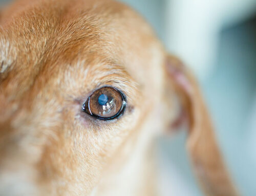
Recognizing the Signs of a Corneal Ulcer
Early Signs
Early detection is key to preserving vision. Watch for these signs:
- Squinting or holding the eye closed
- Excessive tearing or watery discharge
- Cloudiness, haziness, or a bluish tint to the eye
- Rubbing the face or eye on objects
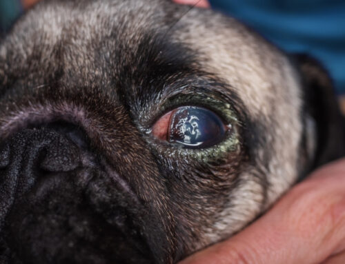
Signs of Worsening
If the ulcer becomes infected or deeper, symptoms may escalate:
- Yellow or green discharge
- Visible white or grey spot on the cornea
- Swelling of the eyelids (blepharedema)
- Reluctance to eat, depression, or other signs of pain
According to the Cornell University College of Veterinary Medicine, any of these symptoms should be taken seriously and evaluated by a veterinarian promptly.
Diagnosing a Corneal Ulcer
In-Clinic Diagnostic Tools
Veterinarians begin by using fluorescein dye, a safe and simple test that highlights any ulcerated area on the cornea. Additional diagnostics may include:
- Slit-lamp exam to assess depth and detect foreign material
- Ocular cytology, as outlined in The Horse’s article on corneal ulcer cytology, to evaluate for fungal or bacterial infection
- Corneal culture and sensitivity testing for non-healing or infected ulcers
For complex or non-responsive cases, referral to a veterinary ophthalmologist is often recommended.
Advanced Diagnostic Imaging
In severe cases or when the eye is too painful to fully examine, your veterinarian may recommend:
- Ocular ultrasound to evaluate for deeper involvement or globe rupture
- Fluorescein retention tests to monitor healing progress over time
Treatment Options for Corneal Ulcers
Medical Management
The treatment plan depends on the depth, size, and presence of infection. Common medications include:
- Topical antibiotics: to prevent or treat bacterial infection
- Antifungals: such as voriconazole or natamycin, especially in humid climates or with fungal risk
- Atropine drops: to dilate the pupil, reduce pain, and prevent adhesions
- Anti-inflammatories: systemic NSAIDs like flunixin or phenylbutazone to reduce inflammation and support comfort
A helpful overview on Equine Stromal Corneal Ulcers is available in this client handout from the University of Tennessee.
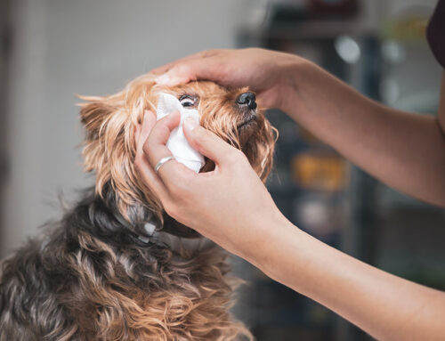
Frequent Medication Is Critical
Corneal ulcers often require frequent dosing—sometimes every 2–4 hours. In cases where topical application is challenging, a subpalpebral lavage system (SPL) can be placed to make administration easier and more consistent.
Surgical Treatment
Some ulcers won’t respond to medical management alone. In these cases, advanced surgical procedures may be necessary, as described by UC Davis Veterinary Medicine:
- Conjunctival grafts to provide support and blood supply to deep ulcers
- Amniotic membrane grafts to promote healing and reduce scarring
- Corneal transplants in select, severe cases
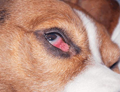
Complications and Prognosis
Potential Complications
Untreated or severe ulcers can lead to:
- Melting ulcers (rapid corneal breakdown from enzymatic activity)
- Corneal perforation
- Uveitis or endophthalmitis (inflammation inside the eye)
- Vision loss or enucleation (removal of the eye in extreme cases)
What Is the Prognosis?
As explained by SmartPak Equine, the outcome depends largely on how early the ulcer is identified and treated:
- Superficial ulcers, when treated early, often heal within 7–10 days with good outcomes.
- Deep, infected, or fungal ulcers have a more guarded prognosis and often require intensive care.
- Prompt intervention dramatically improves the chances of saving the eye and maintaining vision.
Preventing Corneal Ulcers in Horses
You can’t prevent every injury, but some steps help reduce risk:
- Trim long forelocks that trap debris or wick moisture into the eye
- Inspect stalls and paddocks for sharp objects or protrusions
- Use fly masks during turnout, especially in dusty or fly-prone areas
- Clean halters and bridles regularly and inspect for rough spots
- Monitor eye health during trailering or after turnout
Early detection and prompt treatment are your best defenses.
When to Call an Equine Ophthalmologist
Call a veterinary ophthalmologist if:
- The ulcer doesn’t improve within 48 hours
- The horse’s eye becomes more painful
- Discharge becomes purulent (thick, yellow-green)
- The ulcer appears to deepen or spread
- Fungal infection is suspected
At Envision More Veterinary Ophthalmology, we partner with horse owners and referring veterinarians to provide advanced diagnostics and treatments for equine eye conditions, including emergencies.

Why Choose a Veterinary Ophthalmologist?
Board-certified veterinary ophthalmologists bring:
- Specialized equipment to detect and monitor corneal changes
- Advanced medical and surgical treatments not available in general practice
- Extensive experience with equine eye diseases, pain management, and recovery strategies
Our team at Envision More Veterinary Ophthalmology is dedicated to delivering the best possible outcomes for horses with eye conditions.
Corneal ulcers in horses are common but can escalate quickly. Waiting to treat a minor-looking injury can result in significant vision loss—or loss of the eye altogether. The good news? With early intervention and expert care, many horses make a full recovery.
If you suspect your horse has an eye injury, don’t wait. Reach out to your veterinarian or contact our team at Envision More Veterinary Ophthalmology for support.
Leave A Comment